Review Article 
Novel Approach for the Treatment of Cancer: Theranostic Nanomedicine
Mahfoozur Rahman, Mohammad Zaki Ahmad, Imran Kazmi, Sohail Akhter, Yogesh Kumar, Farhan Jalees Ahmad and Firoz Anwar
Pharmacologia, 2012, 3(9), 371-376.
Abstract
Cancer is a growing health problem around the world. At worldwide now more than 10 million cases of cancer per year worldwide. Hence cancer remains one of the leading cause of morbidity and mortality worldwide. Presently major trend in cancer technology research is the targeting of multidrug at a time but it causes drug resistance due to molecular heterogenecity and adoptive resistance in tumor and toxicity. But recently the development of elite nanoparticle which is called as theranostic nanomedicines or multifunctional nanoparticles, it detects the early onset of tumor and same time delivers the drug at a controlled rate. Theranostic nanoparticle has best property due to the magnetic, optical hydrophobicity, higher adsorption capacity, surface charge, specific action and lesser nanotoxicity. Theranostic nanoparticle is the latest technique to diagnose the tumour marker which is involved in the production of tumour. This is also known as all in one system. This review explores the development of elite nanoparticle or theranostic nanomedicines.
ASCI-ID: 5001-68
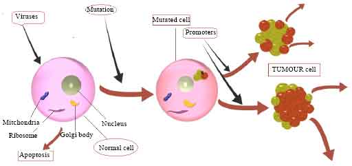
Warner, 2004; Jaffer and Weissleder, 2005). It develops via a multistep carcinogenesis process entailing numerous changes such as cell signalling and apoptosis (Reichert and Wenger, 2008; Zou, 2005). Every cell has normal gene for normal development of cell called as prooncogens, it activation causes formation of oncogene that is responsible for development of abnormal unmatured group of cell called as tumor. Tumor replaces the neighbour healthy cell until tumour reaches to maximum size, moreover healthy cell not be able to compete with tumor cell, resulting ultimately in healthy cell initiate apoptosis. The maximum size of mostly tumor is around 2 mm2. After achieving of maximum size they move to other body part that condition called metastasis which makes cancer incurable (Yezhelyev et al., 2006; Pison et al., 2006). An illustration of tumor development from single cell to maximum size tumor is shown in Fig. 1. Cancer is very complex disease because of molecular heterogenecity (multi phenotype) and adaptive resistance found between tumour cells. Due to this reason it is still the major challenges for its treatment. Conventional treatment for cancer is surgery, radiation, biological therapies (immunotherapy) and chemotherapy. These optional treatments have poor specificity, non recognition of tumour marker, dose related toxicity, poor targeted action, poor bioavailability, neurotoxicity and risk of damage of vital organ (Liong et al., 2008). Thus, we have an urgent need and major opportunities to develop new and innovative technologies that could help to prevent adaptive resistance, identify tumor marker cells and micrometastases. Cancer nanotechnology emerging as a new field of interdisciplinary research, consist of biology, chemistry, engineering and medicine (designing of materials at nanoscale levels to create products (that exhibit novel properties) which have a profound impact on disease prevention, diagnosis and treatment (Ferrari, 2005). In cancer nanotechnology, development of nano based cancer is thergonostic. This is the most effective approach to recognize the molecular heterogenecity and adoptive resistance found in cancer cell.
 |
|
| Fig. 1: | Tumor development from one mutated cell to diffusion-limited
maximum size to metastasis |
 |
|
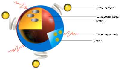
| Fig. 2: | TNM have specific targeting agent, imaging agent,
diagnostic agent, chemotherapeutic agent (drugs), all are in one platform
system |
It reduces the problem associated with conventional therapy in respect of diagnosis, imaging, real time controlled drug release, reduces toxicity and makes a treatment shorter (Tran et al., 2007; Medina et al., 2007; Adiseshaiah et al., 2010; Trochilin, 2005; Gref et al., 1994; Sathe et al., 2006; Dubertret et al., 2002; Pinaud et al., 2004; Lukyanov et al., 2004; Akerman et al., 2002; Farokhzad et al., 2006; Zhu et al., 2007).
Theranostic nanomedicines (TNM)(Table 1), it is an integrated nanotherapeutic system. The term theranostic nanotechnology means combing of individual techniques to form a single nanoplatforms by mounting therapeutic function on them, means combination of diagnostic test with targeted therapy at controlled rate, that co-deliver therapeutic and imaging functions. For successful development of theranostic nanomedicines we must have very well knowledge regarding the metarials science and nanocomposite metarials like particle surface chemistry, non covalent, electrovalent strategies, bio specific interaction hydrophobic adsorption and safety, this will provides better controlled and improved reproducibility of Theranostic nanomedicine (Wang et al., 2001; Huber, 2005). Characteristically it is an amorphous or semi crystalline and colloidal system which has 100-200 nm in size, ideal size range is 10-200 nm, it have better optical, magnetic and thermodynamics properties (Kukowska-Latallo et al., 2005). Efficient in targeting, high drug loading, high specificity on tumour cell or minimize no specificity, improve bioavailability, imaging cell sensitivity, reduce multidrug administration, high spatial resolution, tomographic capability, easily and early onset of detection and deliver selective therapeutic agent (Bai and Wang, 2005).
| Table 1: | Examples of Theranostic nanomedicines |
 |
|
Its function depends on different subunit attached as like anticancer drug, tumour targeting moieties (McCarthy and Weissleder, 2008). These nano based theranostic have four special properties that distinguish them from other cancer therapeutics: (i) the TNM can themselves have therapeutic or diagnostic properties and imaging properties, also it achieve synergistic effects by blocking different receptors (ii) TNM can be attached to multivalent different targeting ligand, it resulting high affinity and specificity for different markers; (iii) TNM can be made to carry multiple drug molecules that simultaneously enable combinatorial cancer therapy and (iv) TNM can bypass traditional drug resistance, molecular heterogeneity and adoptive resistance mechanisms. TNM can achieve increased intracellular concentration by using both passive and active targeting strategies, resulting simultaneously enhancing anticancer effects and reducing systemic toxicity and while minimizing toxicity in normal cells (Acharya et al., 2009).
Several TNM have employed as the carriers of diagnostic agents and drugs (Fig. 2). Lukianova-Hleb et al. (2010) have been studied the optical generation and detection of plasmonic nanobubbles (PNBs) around gold nanoparticles in individual living cells and evaluating the multifunctionality of the PNB. Recently many reviews have discussed about characteristics and biomedical applications of magnetic nanoparticles and mentioned that it can simultaneously act as diagnostic molecular imaging agents and carries different type of drug at same time (Shubayev et al., 2009). Shim et al. (2010) have achieved that coated small-interfering-RNA-encapsulating polyplexes attach with small gold nanoparticles via acid-cleavable linkages to development of combined (theragnostics) stimuli-responsive multimodal optical imaging and stimuli-enhanced gene silencing. This review introduce theranostic agents, in which a linkage between nanoplatforms and functionally entities have been developed and applicable as imaging and therapeutic durg delivering agents.
Cancer molecular imaging: Imaging modality it is a powerful technique to detect primary stage of disease and provide molecular information in cancer (about cellular marker in tumour). Mostly imaging modalities used in drug studies to provide anatomic, pharmacokinetic and pharmacodynamic information. Currently nuclear optical imaging method (PET and single photon emission computed tomography) have superior sensitiveness compared with Computed Tomography (CT), MRI and ultrasound. Right now multimodality imaging is emerging for pharmaceutical application. Imaging co delivered the imaging contrast agent and chemotherapeutic agent which provides real time validation of the targeting strategy (Jaffer and Weissleder, 2005). Optical imaging (Weissleder and Ntziachristos, 2003), utilizes photon emission which is emitted from bioluminescent or fluorescent probes it is less expensive. Photon emission wavelength is visible to near-infrared this provides good resolution (Jain et al., 2008).
Magnetic resonance imaging; It is radiofrequency pulses which is released from water-hydrogen nuclei within an applied magnetic field, higher energy contain nuclei return to the original states this step produces an image. It has good targeted resolution, it requires high concentration to produce a detectible signal and high concentration causes toxicity (Kohler et al., 2006).
Drug delivery in cancer disease: Magnetism engineered iron oxide which conjugate with tumour targeting antibodies, such as trastuzumab which could be used for detecting the tumour cell, cancer cell and release a drug. It is particularly bound to HER2.
HER2 is a tumour marker which is activated in ovarian and breast cancer (Zhang et al., 2005). Iron oxide nanoparticles conjugate with herceptin iron oxide nanoparticles it is used for imaging of tumours in breast cell and also monitor the therapy.
Recently an enzyme guanyl cyclase is presented as a receptor to target a colorectal cancer. Iron oxide based theranostic nanomedicines is best for the imaging and targeting. Release of medicine efficiently (Sakamoto et al., 2005) and also combination of avastin and 5-FU is very useful in colorectal cancers with this conjugation (McBain et al., 2008; Mykhaylyk et al., 2008). In prostrate cancers most commonly flourscien-tagged dendrimer conjugated with specific antibody which is useful in determining the antigen expressing cell with accuracy (Weissleder and Pittet, 2008). Brain cancer is the most lethal type among cancer. Glioma is a tumour and exact measurement of its volume is difficult because of presence of oedema around it. In therapy, problem comes due to the blood brain barrier. For that we formulate a Polysorbate conjugated TNM that is conjugate with peptide which crosses to the blood brain barrier by the mechanism of endocytosis, this mechanism significantly increase the imaging and delivery of therapeutic medicines (Debbage and Jaschke, 2008; Brigger et al., 2002). Recently pancreatic cancer bombesin receptors found in normal acinar cell of pancreas, naturally bombesin which is a peptide obtained from pancreatic cell of European frog Bombina bombina. This conjugate with iron oxide to target a bombesin receptor, that is conjugate particle imaging the tumour cell of pancreatic acinar cell and decrease the intensity of normal pancreatic cell (Storm et al., 1995). Recently latest thergonostic perfluorocarbon-based target has been developed for simultaneously deliver drug molecules as well as ultrasound and MRI contrast agents for various cancers (Tran et al., 2007).
For improving the property of theranostic nanomedicines: For improving the stability and biocompatibility of TNM must attach various group such as polyethylene glycol, modified acrylic acid polymer, phospholipids micelles must attach, that benefit for maintaining drug level in the blood (Bonnemain, 1998; Harisinghani et al., 2003). For improving specific targeting attach aptamer (oligonucleotides), carbohydrates, folic acid and peptides (Senyei et al., 1978; Bonnemain, 1998). For intracellular penetration purpose, we must attach peptides, trans- activating transcriptional activater (TAT) ligand, transferring, positively charged moieties, cationic lipid cationic polymer, resulting pharmacokinetic and bio distribution improves (Harisinghani et al., 2003; Singh et al., 2008). For making the best imaging property we must attach different group to theranostic nanomedicine such as quantum dots, magnetic nanoparticles, it benefits real time nanomedicines biodistributions (Thorek et al., 2006; Yu et al., 2008; Briley-saebo et al., 2004). For making the best stimulus, sensitive drug release property must attach pH labile group photosensitive group, thermosensitive, magnetic sensitive, redox sensitive group. These all provide better control bioavailability and to reduce toxicity (Harisinghani et al., 2003; Pankhurst et al., 2003; Arruebo et al., 2007; Jain et al., 2008). Silica based theranostic nanomedicine applicable for the bio imaging, biosensing and release therapeutic drug. It is a good carrier of metals, drugs and fluoroscent dye its surface can modify and attach different ligand or biomolecules (Dames et al., 2007; Gupta and Gupta 2005; Gupta et al., 2007; Josephson et al., 1999; Zhang et al., 2002; Gref et al., 1994). Moreover, recently Conjugation of silica with dye fluorescien isothiocyanate is use for imaging of human bone marrow stem cell (Bertorelle et al., 2006; Lee et al., 2006; Hamoudeh et al., 2008; Chunfu et al., 2004). Now Core satellite available which is composed of rhodamine dye , silica core and multiple satellite which is made up of magnetic nanoparticles, this combined to humen B1 antibodies which targeting polysialic acid which is a marker of neuroblastoma , lung carcinoma , Wilms’ tumour (Cao et al., 2004). Mesoporous silicate theranostic nanomedicines can conjugate with different ligand and chemo therapeutic drug molecules which are beneficial for diagnosis and treatment of various type of cancer (Liang et al., 2007).
CONCLUSIONS
Theranostic nanomedicines have the potential to target multiple tumor markers and deliver of various therapeutic agents simultaneously in addressing the challenges of adaptive resistance and cancer heterogeneity. This targeted delivery will help to eliminate the need for invasive surgery and radiation therapy. Theranostic medicines improve intra- operative imaging as well as improve pre-operative imaging to visualization of tumours. As the potential of theranostic nanoplatforms continues to increase, the integration of cancer biology to metarials science. In future it will be essential for cancer therapy overall. This novel approach is only effective before tumor cells start metastasizing.
**ad2**
" class="btn btn-success" target="_blank">View Fulltext