Research Article 
Analgesic Effects of an Ethanol Extract of the Fruits of Xylopia aethiopica and Xylopic Acid in Murine Models of Pain: Possible Mechanism(s)
Eric Woode, Elvis O. Ameyaw, George K. Ainooson, Wonder K.M. Abotsi, Eric Boakye-Gyasi and James Oppong Kyekyeku
Pharmacologia, 2013, 4(4), 285-300.
Abstract
Background: The present study examined the possible mechanism(s) of analgesic action of the ethanol extract of the fruits of Xylopia aethiopica (XAE) and its kaurene diterpene constituent, xylopic acid (XA). Methods: Naloxone (2 mg kg-1 i.p.), L-NAME (10 kg-1 i.p.), ondansetron (0.5 mg kg-1 i.p.), theophylline (5 mg kg-1 i.p.), glibenclamide (8 mg kg-1 i.p.), yohimbine (3 mg kg-1 i.p.) and atropine (5 mg kg-1 i.p.) were used to antagonise the anti-nociceptive effects of XAE (100 mg kg-1) and XA (30 mg kg-1) in the formalin test, with morphine as a control. The mechanism was further determined in glutamate-, epinephrine-, capsaicin-, bradykinin- and prostaglandin E2-induced nociception test models. XA was further examined for its μ-opioid receptor (MOR) binding in a [3H]diprenorphine ([3H] DPN) competition binding assay using membranes derived from C6μ cells. Results: The anti-nociceptive effects of XAE, XA and morphine were blocked after naloxone, L-NAME, ondansetron, theophylline and atropine. The effects of XA were also blocked by yohimbine but not glibenclamide. XAE and XA inhibited nociceptive behaviour induced with capsaicin and L-glutamic acid. Additionally, XAE and XA produced anti-hyperalgesic effects in the epinephrine-, bradykinin- and prostaglandin E2-induced hyperalgesia tests. XA and DAMGO were able to competitively displace the binding of [3H]DPN from the MOR but XA appears to bind to two different sites. Conclusion: These results suggest that the anti-nociceptive effects of XAE and XA are mediated through the opioidergic, adenosinergic, muscarinic cholinergic, NO/cGMP, serotoninergic (i.e., via 5-HT3 receptors) pathways. XA additionally acted on the α2- adrenergic system.
ASCI-ID: 5001-157
1. It is one of the largest genera in the Annonaceae and belongs to the subfamily Annonoideae, tribe Unoneae and subtribe Xylopiineae2. Xylopia aethiopica (Dunnal) A. Rich, one of the species widely distributed in Ghana, is a slim, tall, evergreen, aromatic tree that grows up to 15-30 m high. The fruit, commonly known as African pepper or locally as Hwentia (Twi), Tso (Ewe) and Soo (Ga), is as a spice in the preparation of soup. In Ghanaian traditional medicine, the fruit extract is used for the treatment of cough, rheumatism, lumbago, headache, neuralgia and colic pain3,4. It was employed in government hospitals in Ghana to induce placental discharge postpartum due to its abortifacient effect5,6.
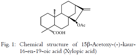
Previous studies on various extracts of the fruits and seeds have showed antimicrobial activity7,8, cytotoxic activity8,9, hypotensive and diuretic activity10, termite antifeedant activity11, intraocular pressure lowering effect6, hypolipidaemic and antioxidant activities12,13. Phytochemical constituents in the fruit and seeds of X. aethiopica include diterpenic acids (xylopic acid, kaurenoic acid, 15-oxo-kaurenoic acid, etc), diterpenic alcohols (kauran-16-α-ol, etc), acyclic compounds, essential oils, volatile oils, alkaloids, glycosides, saponins, tannins, sterols and cuminal5,14,15,16. Xylopic acid [15β-acetoxy-(-)-kaur-16-en-19-oic acid] (Fig. 1) and kaurenoic acid (ent-kaur-16-en-19-oic acid), the major constituents of Xylopia aethiopica, are reported to exhibit a number of biological activities including antimicrobial7,17, cytotoxic18,19, anti-HIV20, hypotensive and diuretic10, anti-inflammatory and antipyretic21 activities.
We have earlier reported the analgesic properties of the ethanol extract of the fruits of Xylopia aethiopica (XAE) as well as its kaurene diterpene constituent, xylopic acid (XA)22.
 |
In this study, we examined further, the analgesic activities of XAE and XA in an attempt to elucidate their possible mechanism(s) of action.
MATERIALS AND METHODS
Collection of plant material: The dried fruits of Xylopia aethiopica were collected from the Botanical Gardens (06°41’6.39"N; 01°33’45.35"W) of Kwame Nkrumah University of Science and Technology (KNUST), Kumasi, Ghana between the months of August and December, 2008. The fruits were authenticated by Dr. Kofi Annan at the Department of Pharmacognosy, Faculty of Pharmacy and Pharmaceutical Sciences, College of Health Sciences, KNUST. A voucher specimen (No. FP/09/77) has been kept at the herbarium of the Faculty.
Preparation of the ethanol extract of Xylopia aethiopica (XAE): A quantity of the fruit was pulverized into fine powder. About 0.36 kg of the powdered material was placed in cylindrical jars and macerated with 70% (v/v) ethanol for three days. The filtrate was concentrated using rotary evaporator at a temperature of 60°C. This resulted in a greenish solid mass of ethanol extract of Xylopia aethiopica with a percentage yield of 34.8% (w/w).
Isolation and purification of xylopic acid (15β-Acetoxy-(-) - kaur-16-en-19-oic Acid): The extraction and purification process was same as what was described by Woode et al.22. The yield of the isolated/purified xylopic acid (Fig. 1) was 1.41% (w/w). Purity of the isolated xylopic acid was determined using high performance liquid chromatography (HPLC) as described by Adosraku and Kyekyeku23. The chromatograph consisted of LC-10AT Shimadzu pump with programmable absorbance detector (783A Applied Biosystems) and Shimadzu CR501 Chromatopac. Phenomenex Hypersil 20 micron C18 200x3.20 mm column was used. The mobile phase consisted of methanol and water (9:1) eluted isocratically at 0.5 mL min-1. Portions of 20 μL of a suitable concentration of pure XA were loaded and injected unto the column after dissolving in the mobile phase at 60°C. The eluent was monitored at 206 nm. Portions of the XAE and XA were loaded and injected. The peak(s) were noted as component(s) of the XAE and XA.
Drugs and chemicals: The drugs and chemicals used were: Naloxone, NG-Nitro-l-arginine methyl ester/L-NAME (Troge Medical Gmbh, Hamburg, Germany); morphine hydrochloride (Phyto-Riker, Accra, Ghana); carrageenan, L-glutamic acid, prostaglandin E2, capsaicin, bradykinin acetate, DAMGO (Sigma-Aldrich Inc., St. Louis, MO, USA), glibenclamide (Daonil®, Sanofi-Aventis, Guildford, UK); formalin, theophylline (BDH, Poole, England); Ondansetron (GlaxoSmithKline, Uxbridge, UK); Atropine sulphate (E. Merck AG-Darmstadt, Germany); yohimbine (Walter Ritter Gmbh + Co. KG, Germany); epinephrine HCl (Wuhan Grand Pharm, China); ketamine HCl (Brotex Medica, Trittau, Germany); propranolol (Ernest Chemist Ltd, Accra, Ghana); captopril (Teva UK Ltd, Eastbourne, UK); Tissue culture media, Geneticin, Fetal bovine serum (Invitrogen, Carlsbad, CA); [3H]diprenorphine (Perkin-Elmer Life Sciences, Boston, MA) and Ecolume scintillation fluid (ICN, Aurora, OH). All other biochemicals used in the binding studies were obtained from Sigma-Aldrich Inc. (St. Louis, MO, USA). All drugs used in the nociceptive tests were dissolved in normal saline except for capsaicin which was dissolved in 0.5% ethanol. The ethanol did not cause an effect “per se”.
Animals: Sprague-Dawley rats (250-300 g) and ICR mice (20-25 g) of both sexes were purchased from Noguchi Memorial Institute for Medical Research, University of Ghana, Legon, Ghana and housed in the animal facility of the Department of Pharmacology, Kwame Nkrumah University of Science and Technology (KNUST). The animals were housed in groups of six in stainless steel cages (34x47x18 cm) with soft wood shavings as bedding, fed with normal commercial pellet diet (GAFCO, Tema), given water ad libitum and maintained under laboratory conditions (temperature 24-25°C, 12 h light-dark cycle). All procedures and techniques used in these studies were in accordance with the National Institute of Health Guidelines for the Care and Use of Laboratory Animals (NIH Publication No. 85 - 23, 1985, revised 1996). All protocols used were approved by the Departmental Ethics Committee.
Glutamate-induced nociception: The possible interaction of XAE and XA with the glutamatergic system was assessed with the glutamate test. The procedure used was similar to that described by Woode and Abotsi24. Male mice were pre-treated with XAE (30-300 mg kg-1, p.o.), XA (10-100 mg kg-1, p.o.) or ketamine (1-10 mg kg-1, i.p.) or vehicle (normal saline, 10 mL kg-1, i.p.) 30 min (i.p.) or 1 h (p.o.) before intraplantar injection of glutamate (10 μmoL paw-1, 20 μL). Data were expressed as the Mean±SEM of nociceptive scores between 0 and 15 min after glutamate injection.
Capsaicin-induced nociception: The test was performed as described by Woode and Abotsi24. Male mice were pre-treated with XAE, XA or vehicle similar to that described for the glutamate test above, 30 min (i.p.) or 1 h (p.o.) before intraplantar injection of capsaicin (1.6 μg paw-1, 20 μL dissolved in 0.5% ethanol). The amount of time spent licking the injected paw was determined for 5 min following capsaicin injection and was considered as a nociceptive behaviour.
Bradykinin-, prostaglandin E2- and epinephrine-induced hyperalgesia: The procedure used was similar to that described by Meotti25. After baseline pain threshold measurements, rats (males) were treated with XAE (30-300 mg kg-1, p.o.), XA (10-100 mg kg-1, p.o.), or vehicle 30 min (i.p.) or 60 min (p.o) before the intraplantar injection of bradykinin (10 nmoL paw-1), prostaglandin E2 (1 nmoL paw-1), epinephrine (450 nmol paw-1) or normal saline. Morphine (1-10 mg kg-1, i.p.) and propranolol (1-10 μg paw-1) were used as reference drugs in prostaglandin E2- and epinephrine-induced hyperalgesia, respectively. Mechanical nociceptive thresholds were measured in the rat paw pressure test using an analgesimeter (IITC Life Science Model 2888, Woodland Hills, CA, USA) which is based on the Randall and Sellito test26. This was used to apply a linearly-increasing pressure, by means of a blunt perspex cone, to the dorsal region of the right hind paw until the rat withdrew the paw. Rats received two training seasons before the day of testing. Pressure was gradually applied to the right hind paw and Paw Withdrawal Thresholds (PWTs) were assessed as the pressure (grams) that elicits paw withdrawal. A cut-off point of 250 g was used to prevent any tissue damage to the paw. A change in hyperalgesic state was calculated as a percentage of the maximum possible effect (% MPE). Rats were pre-treated with captopril (an angiotensin-converting enzyme inhibitor; 5 mg kg-1, s.c.) 1 h before experiments to prevent the degradation of bradykinin.
Further assessment of mechanisms in the formalin test: The mechanism of analgesic action of XAE and XA was further investigated using various antagonists in the formalin test. The doses of antagonist, agonist and other drugs were selected on the basis of previous literature data and in pilot experiments in our laboratory24,27. In all these experiments, animals received XA (30 mg kg-1, p.o.), XAE (100 mg kg-1, p.o.), morphine (3 mg kg-1, i.p.) or vehicle 15 min after administration of the antagonist except animals pre-treated with yohimbine or glibenclamide, in which case XAE, XA or morphine was administered after 30 min. The nociceptive response to the formalin injection was recorded 60 min after administration of XAE, XA or vehicle and 30 min after administration of morphine.
To assess the involvement of the opioid system, mice were pre-treated intraperitoneally with naloxone (a non-selective opioid receptor antagonist; 2 mg kg-1) or vehicle. For the adenosinergic system, mice were pre-treated with theophylline (a non-selective adenosine receptor antagonist; 5 mg kg-1, i.p.) or vehicle. To investigate the involvement of ATP-sensitive K+ channels, mice were pre-treated with glibenclamide (an ATP-sensitive K+ channel inhibitor; 8 mg kg-1, p.o.) or vehicle. Assessment of the possible involvement of nitric oxide in the anti-nociceptive effects of XA and XAE was evaluated by pre-treating mice intraperitoneally with Nù-Nitro-L-arginine methyl ester hydrochloride/L-NAME (a NO synthase inhibitor; 10 mg kg-1, i.p.) or vehicle. To assess the possible involvement of 5-HT3 receptors in the anti-nociceptive effects of XA and XAE, mice were pre-treated intraperitoneally with ondansetron (5-HT3 receptor inhibitor, 0.5 mg kg-1) or vehicle. For the muscarinic cholinergic system, mice were pre-treated with 5 mg kg-1 i.p. atropine (non-selective muscarinic antagonist) or vehicle. The possible contribution of α2-adrenoceptors to the anti-nociceptive effects of XA and XAE was determined by pre-treating mice intraperitoneally with yohimbine (an α2 receptor antagonist; 3 mg kg-1) or vehicle.
Radioligand binding assay: Xylopic acid was examined for its μ-opioid receptor (MOR) binding in a [3H]diprenorphine ([3H] DPN) competition binding assay using membranes derived from C6μ cells28.
Cell culture: C6 glioma cells stably transfected with the u-opioid receptor (C6μ) were maintained at 37°C under 95% O2/5% CO2 in media (DMEM) containing 10% Fetal Bovine Serum (FBS) and 500 μg mL-1 Geneticin® (Invitrogen) to select for the u-opioid receptor-expressing cells. Cells were grown to in 10 cm2 dishes to ~90% confluency on the day of the assay.
Membrane preparation: C6μ cells were washed twice with ice cold phosphate-buffered saline (0.9% NaCl, 0.61 mM Na2HPO4 and 0.38 mM KH2PO4, pH 7.4), detached from the plate by incubation in warm harvesting buffer (20 mM HEPES, pH 7.4, 150 mM NaCl and 0.68 mM EDTA) for 5 min, dispersed by agitation and pelleted by centrifugation at 1600 rpm for 3 min. The resulting cell pellet was resuspended in ice cold 50 mM Tris buffer, pH 7.4 and homogenized with a Tissue Tearor (Biospec Products Inc., Bartlesville, OK) for 20 sec. The homogenate was centrifuged at 25000 xg at 4°C for 20 min. The resulting membrane pellet was resuspended in 50 mM Tris buffer, pH 7.4 and centrifuged as above. The final pellet was resuspended in 50 mM Tris, pH 7.4 with a Dounce homogenizer, and the homogenate was separated into 0.5 ml aliquots and stored at -80°C until use. Protein concentration was measured by the Bradford protein assay using bovine serum albumin as standard29.
Competition binding assay: For competition binding assays, cell membranes (15 μg protein) were incubated at room temperature (25°C) for 1 h with shaking with 0.2 nM [3H]diprenorphine ([3H]DPN) and increasing concentrations of unlabeled ligand [xylopic acid (0.1nM - 1 mM) or DAMGO (0.001nM - 10 μM)] in 50 mM Tris-HCl, pH 7.4. Nonspecific binding was determined in the presence of 10 μM naloxone. The assay was stopped by rapid filtration through glass GF/C filters (Whatman, Clifton, NJ) using a Brandel harvester (MLR-24, Gaithersburg, MD), rinsing three times with ice-cold 50 mM Tris buffer, pH 7.4. Filters were dried at 50°C for 20 min. Dried filters were saturated with EcoLume liquid scintillation mixture (MP Biomedicals, Solon, OH), heat sealed in polyethylene bags and radioactivity was counted in a Wallac 1450 MicroBeta Liquid Scintillation and Luminescence Counter (Perkin Elmer, Boston, MA).
Data analysis: GraphPad Prism for Windows version 4.03 (GraphPad Software, San Diego, CA, USA) was used for all data and statistical analyses. The p<0.05 was considered statistically significant. In all nociceptive tests, a sample size of seven animals (n = 7) were used. The time-course curves were subjected to two-way (treatmentxtime) repeated measures Analysis of Variance (ANOVA) with Bonferroni’s post hoc test. Total nociceptive score for each treatment was calculated in arbitrary unit as the area under the curve (AUC). Differences in AUCs were analysed by ANOVA followed by Student-Newman-Keuls post hoc test. Doses for 50% of the maximal effect (ED50) for each drug were determined by using an iterative computer least squares method, with the following nonlinear regression (three-parameter logistic) equation:
 |
where, X is the logarithm of dose and Y is the response. Y starts at a (the bottom) and goes to b (the top) with a sigmoid shape.
The fitted midpoints (ED50s) of the curves were compared statistically using F test30,31.
In the binding studies, each data point represents the Mean±SEM of duplicate wells of at least two experiments. Specific binding of [3H]DPN was determined by subtracting non-specific binding counts from all values and data were expressed graphically as a percentage of the total [3H]DPN binding ([3H]DPN binding in the absence of unlabeled ligand) against increasing concentration of unlabeled ligand (xylopic acid or DAMGO). Binding data were fit by non-linear regression using one- or two-site binding models and when possible the affinity of xylopic acid or DAMGO binding to the μ-opioid receptor (Ki or IC50) was determined from the resulting curve fit. The IC50 was defined as the concentration of drug that inhibited the specific [3H]diprenorphine binding by 50%.
RESULTS
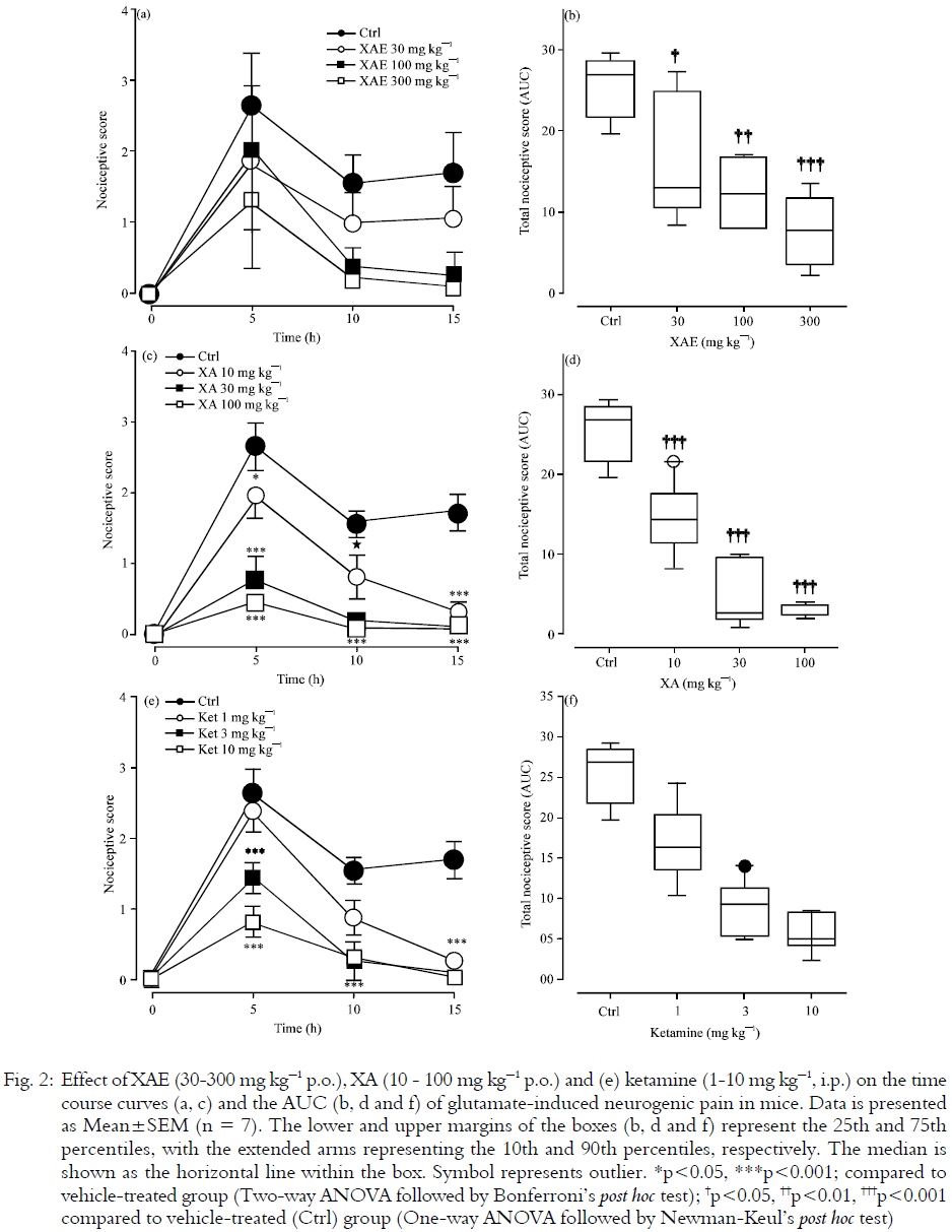
Glutamate-induced nociception: Intraplantar administration of glutamate induced nociceptive behaviour (biting, flinching and licking of the injected paw) in mice during the 15 min period of observation. Treatment of mice with XAE (30-300 mg kg-1, p.o.) produced dose-dependent inhibition of glutamate-induced neurogenic nociception (F3,24=10.02, P=0.0002, Fig. 2a) with a maximum effect of 69.60±2.1% at the highest dose (Fig. 2b). XA (10-100 mg kg-1, p.o.) also produced marked, dose-related inhibition of glutamate-induced neurogenic pain (F3,24= 32.52, P<0.0001; Fig. 2c) with the highest dose causing a maximum anti-nociception of 88.9±0.31% (Fig. 1d). The reference drug, ketamine (1-10 mg kg-1, i.p.), inhibited glutamate-induced nociception in a dose-related manner (F3,24= 24.31, p<0.0001; Fig. 2e). Ketamine reduced the nociception evoked by glutamate by 77.4±1.20% at 10 mg kg-1 (Fig. 2f).
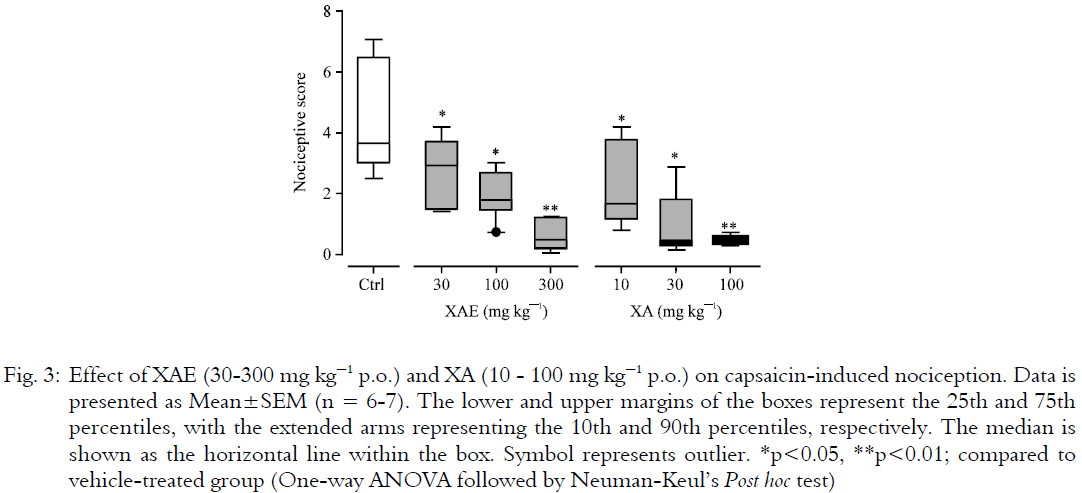
Capsaicin-induced neurogenic pain: Intraplantar injection of capsaicin in to mice produced biting and licking response toward the injected paw. This started immediately after the administration of capsaicin and diminished after 5 min. Oral treatment of the animals with XAE (30–300 mg kg-1) produced profound, dose-related anti-nociception (F3,24=6.84, p=0.0017; Fig. 3) with a peak effect of 86.0±0.23% at 300 mg kg-1(Fig. 3). XA (10–100 mg kg-1, p.o.) also produced dose-related attenuation of capsaicin-induced neurogenic nociception (F3,23=6.65, p=0.0021; Fig. 3) with maximal effect of 89.10±0.1% at the highest dose (Fig. 3). The rank order of efficacy was XAE>XA. The opposite was true for their potencies (Table 1).
 |
 |
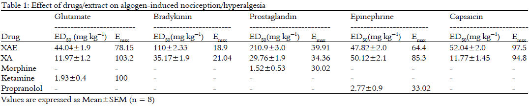 |
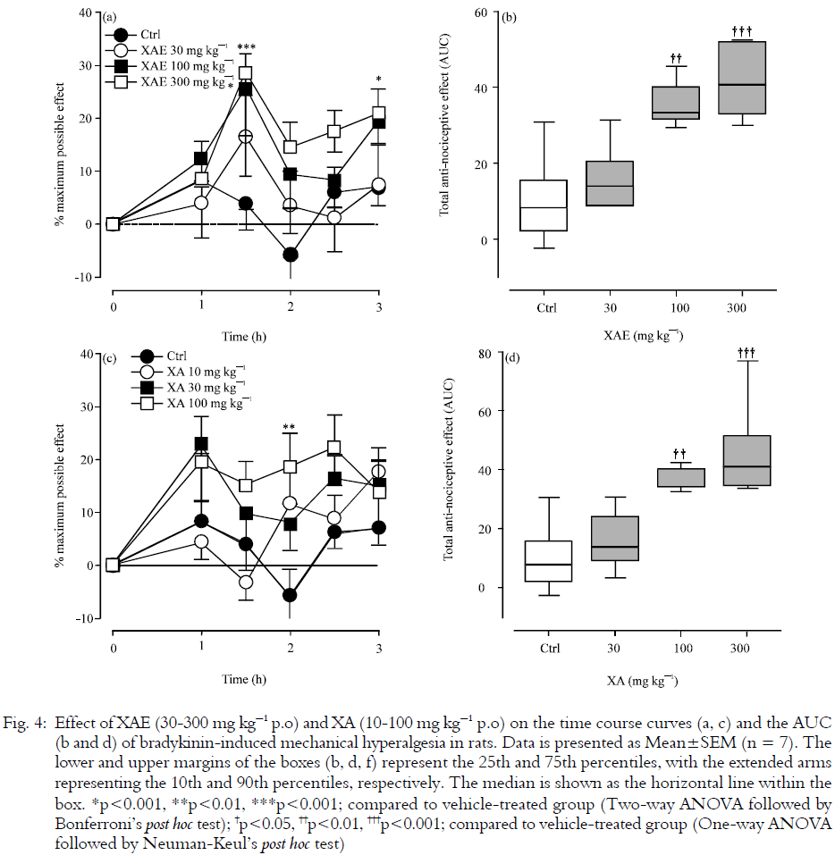
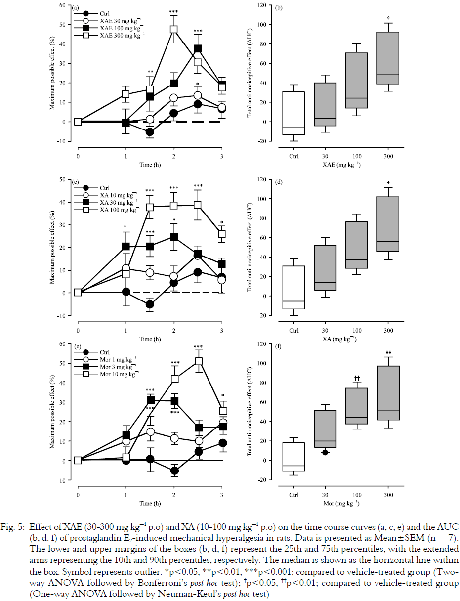
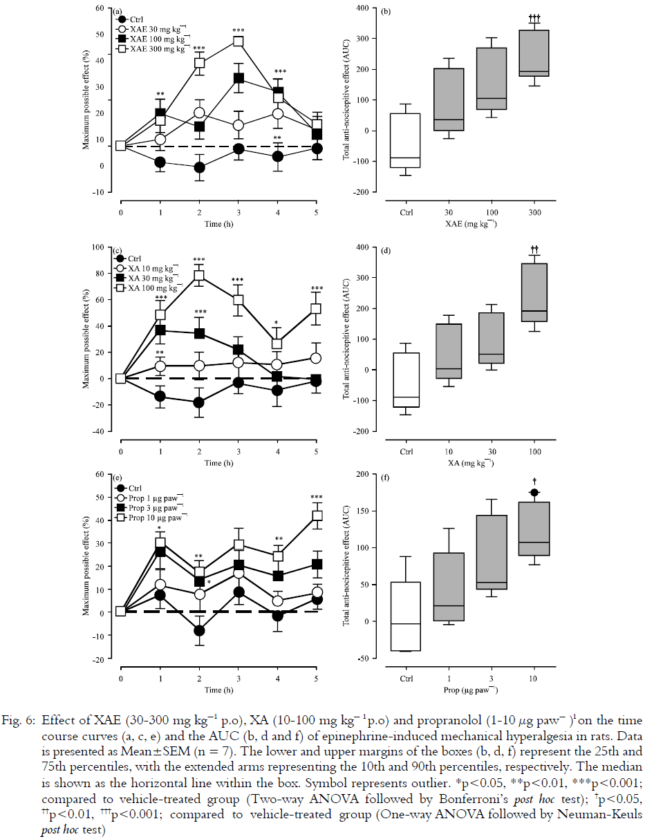
Bradykinin-, prostaglandin E2- and epinephrine-induced hyperalgesia: Figures 4, 5 and 6 show the results obtained in the hyperalgesia tests. Mechanical hyperalgesia was induced in all the animals treated with bradykinin, PGE2 or epinephrine although the control group exhibited higher hyperalgesia compared to the treated groups. Administration of XAE (30-300 mg kg-1, p.o.) significantly and dose-dependently inhibited bradykinin- (F3,24 = 8.85, p=0.0004), PGE2- (F3,24 = 3.72, p = 0.0249) and epinephrine-induced (F3,24 = 4.97, p = 0.008) mechanical hyperalgesia in rats with maximal effect at 300 mg kg-1. Similarly, oral administration of XA (10–100 mg kg-1) also significantly attenuated bradykinin- (F3,24 = 8.23, p = 0.0006), PGE2- (F3,24 = 5.0, p = 0.0078) and epinephrine-induced (F3,24 = 5.83, p = 0.0039) mechanical hyperalgesia in a dose-dependent manner with the highest inhibition at 100 mg kg-1. Morphine (1-10 mg kg-1) used as reference drug in the PGE2-induced hyperalgesia test also produced significant (F3,24 = 6.45, p = 0.0023), dose-related reduction in hyperalgesia while propranolol (1–10 μg paw-1) showed similar effects (F3,24 = 4.62, p = 0.0109) in the epinephrine test. XA was more efficacious than XAE in all the tests (Table 1).
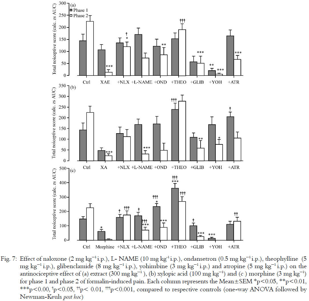
Analysis of mechanisms in the formalin test: Figure 7 shows the effect of naloxone, glibenclamide, L-NAME, yohimbine, atropine, theophylline and ondansetron on the anti-nociceptive effects of XAE (100 mg kg-1, p.o.), XA (30 mg kg-1, p.o.) and morphine (3 mg kg-1, i.p.). Pre-treatment of mice with naloxone (2 mg kg-1, i.p.) and theophylline (5 mg kg-1, i.p.) reversed the analgesic effects of XAE, XA and morphine in both phases of the formalin test.
L-NAME (10 mg kg-1, i.p.) and atropine (5 mg kg-1, i.p.) blocked the neurogenic (Phase 1) anti-nociception of XAE and XA (Fig. 7a, b). L-NAME also blocked the anti-nociception of morphine (3 mg kg-1, i.p.) in both phases of the formalin test while atropine blocked it only in the second (Fig. 7c).
Pre-treatment of the animals with ondansetron (0.5 mg kg-1) blocked the second phase of XAE-induced anti-nociception (Fig. 7a). Ondansetron also antagonized XA- and morphine-induced anti-nociception in the first phase of the formalin test (Fig. 7b, c).
Also, yohimbine (3 mg kg-1) reversed the neurogenic anti-nociception of XA (Fig. 7b) but had no effect on analgesic activity of XAE and morphine (Fig. 7a,c) in both phases of formalin-induced pain.
 |
Furthermore, glibenclamide (8 mg kg-1, i.p.), partially reversed the anti-nociception of morphine in the first phase but failed to significantly block the anti-nociception of XAE and XA in both phases of the formalin test.
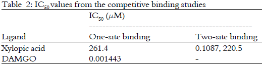
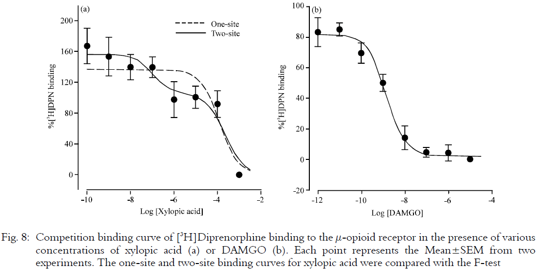
Radioligand binding assay: Data for DAMGO was best fit by a single binding site model. However, a comparison of the one-site/two-site model curve fits of the data for xylopic acid revealed that the two-site model was better [F1,12= 5.533, P= 0.0366; R2 (one-site) = 0.74, R2 (two-site) = 0.82]. Xylopic acid and DAMGO were able to competitively displace the binding of [3H]DPN from the MOR (Fig. 8). Table 2 shows the IC50 values of the inhibitions. XA appears to bind to two different sites with IC50 values of 0.11 and 220.5 μM, respectively. DAMGO showed very high affinity to the MOR with IC50 and Ki values of 0.0014 μM and 0.47 nM, respectively. The affinity of DAMGO (based on the IC50 values) is about 100 times greater than XA at its high affinity binding site.
 |
 |
 |
 |
There was also an apparent increase in [3H]DPN binding that occurred at low concentrations of xylopic acid (Fig. 8a).
**ad4**
DISCUSSION
This study is a follow-up on our earlier study22 in which we showed that the ethanol extract of the fruits of Xylopia aethiopica (XAE) as well as its kaurene diterpene constituent, xylopic acid (XA) have analgesic properties in animal models of acute, persistent and peritoneovisceral pain. Our findings, in the current study, strongly indicate that the analgesic effects of XAE involve an interaction with the opioidergic, adenosinergic, muscarinic cholinergic, NO/cGMP and serotonergic systems. The mechanisms for XA are similar but additionally involve α2-adrenergic system.
Nociception involves a complex interaction of peripheral and central nervous system structures, extending from the skin, the viscera and the musculoskeletal tissues to the cerebral cortex.
 |
Several mediators-neurotransmitters, ion channels, receptors, and second-messenger systems of nociceptive neurons-work together at a molecular level to ensure the generation, processing and transmission of nociceptive information which helps to prevent tissue damage.
In order to identify some possible mediators involved in the anti-nociceptive activity of XAE and XA, an antagonism study was carried out in the formalin test. The anti-nociceptive effect of XAE and XA was assessed in the presence of various antagonists including naloxone, theophylline, L-NAME, glibenclamide, atropine, ondansetron and yohimbine. All the antagonists used did not produce anti-nociception when administered alone. Systemic administration of the opioid receptor antagonist naloxone inhibited the anti-nociceptive effects of XAE and XA in both phases of the formalin test. This strongly suggests an opioidergic involvement in the actions of XAE and XA.
Our results also show that pre-treatment of the mice with NG-Nitro-L-arginine methyl ester (L-NAME; NO synthase inhibitor) reversed the anti-nociception of XAE and XA in the neurogenic phase of the formalin test. This finding implicates the involvement of the NO/cGMP pathway. Nitric oxide plays a complex and diverse role in the modulation of nociceptive transmission in both the peripheral and central nervous system32. NO exerts a dual effect on nociception and there are suggestions that this may be due to the existence of different subsets of nociceptive primary sensory neurons in which NO plays opposing roles32,33. ATP-sensitive K+ channels seem not to be involved in the actions of XAE and XA since glibenclamide (ATP-sensitive K+ channel blocker) did not significantly alter their anti-nociceptive effects. Potassium channels other than the ATP sensitive K+ channels involvement may not entirely be excluded.
The anti-nociceptive effects of XAE and XA were reversed by pre-administration of theophylline implicating the involvement of adenosinergic pathway in their actions. Adenosine acts at several P1 receptors (A1, A2A, A2B, and A3) all of which are coupled to G proteins34. In the periphery, adenosine A1 receptor activation produces pain suppression, while adenosine A2 receptor activation produces pain enhancement35. Within the spinal cord, adenosine A1 receptor activation produces anti-nociception35. Adenosine A3 receptor activation produces pro-nociception in the CNS35 and also peripherally-secondary to mast cell degranulation and release of histamine and 5-hydroxytryptamine (5-HT) that exert nociceptive actions at sensory nerve terminal35. Since theophylline blocks adenosine A1 and A2 receptors, the anti-nociceptive effects may be due to activation of A1 receptors and/or an increment in endogenous adenosine either centrally or peripherally. The finding that both opioidergic and adenosinergic mechanisms are possibly involved in the anti-nociceptive effects of XAE and XA is not surprising since both opioid and adenosine receptor agonists are known to share some similar anti-nociceptive mechanisms36,37. The A1 receptor has been proposed to exist as part of a multireceptor complex, in association with μ-opioid and α2-adrenergic receptors on the basis of a demonstrated cross antagonism, cross tolerance and cross withdrawal between these systems35,38 and activation of one of these receptors may affect the rest39. The spinal release of adenosine contributes to the anti-nociceptive activity of μ-opioid agonists40,41. This explains why theophylline inhibited the anti-nociception of morphine in this study.
Activation of muscarinic receptors induces anti-nociception in various pain paradigms including thermal, inflammatory and neuropathic pain42,43. M1, M2, M3 and M4 receptors are involved in mediating the analgesic effects of muscarinic agonists at the spinal and supraspinal level42, 44,45,46,47 while peripheral activation of M2 receptors likely contributes to analgesia via reduced CGRP release42,48. Our results show that anti-nociceptive effects of XAE and XA involve the muscarinic cholinergic pathway since it was antagonized by atropine. Atropine also blocked the analgesic effects of morphine. It is already well established that the analgesic effect of systemic morphine is mediated by a descending cholinergic pathway49 as well as spinal endogenous acetylcholine acting through muscarinic receptors50,51,52,53.
Pre-treatment of mice with yohimbine (an α2-adrenoceptor antagonist) also reversed the anti-nociception of XA in the first phase of the formalin test but affected neither XAE nor morphine. This implicates the involvement of α2-adrenergic mechanisms in the anti-nociceptive activity of XA. Alpha-2-receptors are important in peripheral, spinal and supraspinal pain modulation. In the spinal cord, for instance, norepinephrine released from descending pathways suppresses pain by inhibitory action on α2A-adrenoceptors on central terminals of primary afferent nociceptors (presynaptic inhibition) or by direct α2-adrenergic action on pain-relay neurons (postsynaptic inhibition)54. Serotoninergic neurons also play a crucial role in the control of pain55 and the diversity of subtype receptors for serotonin, makes this system able to exert either facilitatory or inhibitory function56. Spinal 5-HT3 receptors haven been shown to mediate anti-nociception, possibly via GABA release57,58. Pre-administration of ondansetron attenuated the anti-nociceptive of XAE and XA in the formalin test. This implicates 5-HT3-serotoninergic involvement in the anti-nociceptive activity of XAE and XA.
The μ-receptor subtype (MOR) is generally thought to be responsible for most of the analgesic effects of the opioids. Since antagonism in the formalin test revealed the involvement of opioidergic pathways, our study investigated if XA actually binds to opioid receptors via radioligand competitive binding studies. Considering, the IC50 values of XA in comparison with DAMGO, it appears that XA has some (albeit moderately weak) affinity for MOR. Unfortunately, it is not yet clear what the binding sites actually represent-it could signify binding to more than one of the proposed μ-opioid receptor subtypes58,59 or it could represent both low and high affinity binding to the same μ-receptor (e.g., high affinity in the presence of G protein, low affinity in the absence). The involvement of other opioid receptor subtypes (i.e., δ or κ) in the analgesic actions of XA is also possible and this requires further investigation. Perhaps, the most interesting finding from the binding studies was the apparent increase in [3H]DPN binding that occurred at very low concentrations of XA. It suggests that XA may be able to enhance the binding of orthosteric ligands, such as DPN. Xylopic acid may, therefore, be producing its analgesic effects by acting as an allosteric modulator of the μ-opioid receptor in vivo to potentiate the anti-nociceptive effects of endogenous μ-opioid receptor ligands. Further studies are necessary to confirm this observation.
In order to ascertain the involvement of the glutamatergic system in the mechanism of action of XAE and XA, the glutamate test was carried out with ketamine as a reference drug. XAE, XA and ketamine attenuated the nociceptive behaviour induced by glutamate. Beirith et al.60 have shown that the nociceptive response induced by glutamate appears to involve peripheral, spinal and supraspinal sites of action and is greatly mediated by both NMDA and non-NMDA receptors by a mechanism which largely depends on the activation of L-arginine-nitric oxide pathway. The anti-nociceptive activity of XAE and XA in the glutamate test, therefore, probably involves an inhibition of the production/action of NO, or an interaction with the glutamatergic system. This is a significant finding because glutamate and its receptors, both ionotropic and metabotropic, are critical for nociceptive processing. Hyperfunction or dysfunction of glutamatergic neurotransmission is known to be a key mechanism of pain-related plastic changes in the central and peripheral nervous system61. NMDA receptor-mediated mechanisms are particularly important as NMDA-receptor antagonists have been shown to effectively alleviate pain-related behavior, reduce opioid-induced hyperalgesia and retard opioid tolerance development in both animal models as well as in clinical situations62,63,64.
Our results also demonstrate that XAE and XA suppressed nocifensive responses in the capsaicin test. Capsaicin activates the vanilloid receptor, TRPV1, a ligand-gated non-selective cation channel present in primary sensory neurons65. This leads to the release of neuropeptides, excitatory amino acids, nitric oxide and pro-inflammatory mediators from the periphery and transmits nociceptive information to the spinal cord or causes spinal sensitization through protein kinase A and C activation25,66. The anti-nociceptive effect of XAE and XA may therefore involve an interaction with TRPV1 or the inhibition of production or action of some of these mediators. It has been shown that μ-opioid receptor activation can inhibit the activity of TRPV1 via Gi/o proteins and the cAMP pathway67. Therefore, XA (and most likely XAE) may have acted to inhibit capsaicin-induced nociception through the opioid pathway.
This study has also revealed that XAE and XA inhibit bradykinin-, prostaglandin E2 -and epinephrine-induced hyperalgesia. Bradykinin, an important peripheral mediator of pain, elicits nociception or hyperalgesia by direct stimulation of the nociceptors Aδ and C-fibers. Several inflammatory and algogenic substances such as products derived from arachidonic acid pathways, cytokines and nitric oxide as well as neuropeptides such as calcitonin gene-related peptide and substance P may also be released by bradykinin68,69. The mechanical hyperalgesia induced by bradykinin involves a B2 receptor-mediated direct activation of protein kinase C and the indirect activation of the protein kinase A25,70. Epinephrine and prostaglandin E2 also act through β and EP receptors respectively, to activate cAMP/PKA and PKC pathways leading to hyperalgesia71,72. It is speculated that XA and XAE may have acted directly or indirectly to inhibit B2/β/EP receptors or PKA and/or PKC pathways.
CONCLUSION
In conclusion, our study has revealed that the analgesic effects of XAE involve an interaction with the opioid, adenosinergic, muscarinic cholinergic, NO/cGMP and serotonergic systems (i.e., through 5-HT3 receptors). The mechanisms for XA are similar but additionally involve α2-adrenergic receptors.
ACKNOWLEDGMENTS
We wish to thank Prof. John Traynor (PhD) and Jenny Lamberts (Graduate Student) of the Department of Pharmacology University of Michigan, Ann Arbor, MI for their assistance in the radioligand binding studies. We are also grateful to Ghana-Michigan CHARTER Research Fellowship Programme. George Kwaw Ainooson was sponsored by the Ghana-Michigan CHARTER Research Fellowship Programme. We are also grateful to all the Laboratory Technicians of the Department of Pharmacology especially Mr. Thomas Ansah.
" class="btn btn-success" target="_blank">View Fulltext